Allgemein
Hauptmerkmale
- Glisteningfreies hydrophobes Acrylat ¹, ², ³, ⁴, ⁵, ⁶, ⁷, ⁸, ⁹, ¹⁰
- Die texturierte Optikkante wurde konzipiert, um Dysphotopsien zu reduzieren
- Reduziertes PCO-Risiko (Posterior Capsular Opacification) durch scharfes Kantendesign und innovative Oberflächenbehandlung
- Texturiert raue Haptiken
- Aussendurchmesser der vorderen Injektorspitze 1.70 mm
- Langzeit-Transparenz basierend auf In-vitro-Tests ⁹
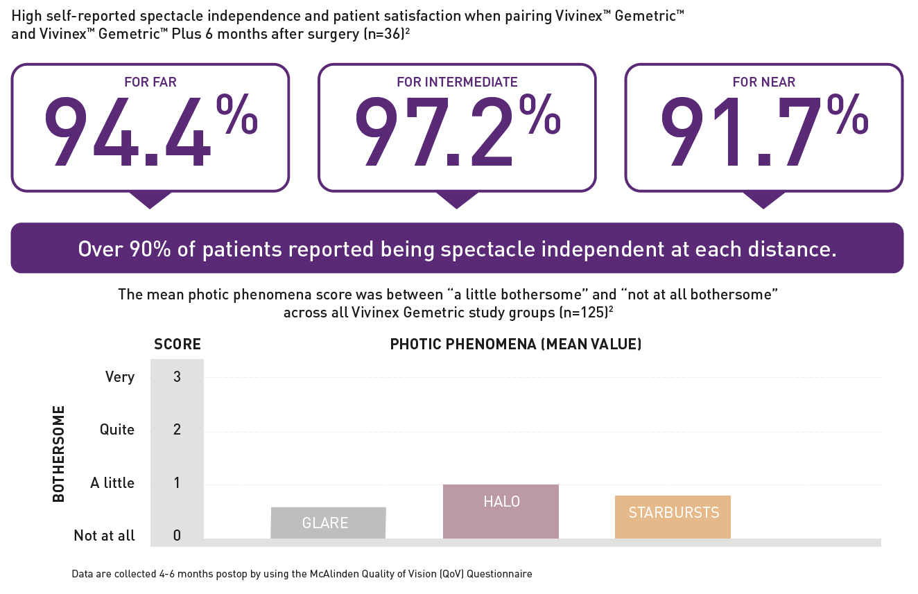
- Hohe Brillenfreiheit von über 90 % der Patienten ¹
Der Weg zur Brillenunabhängigkeit
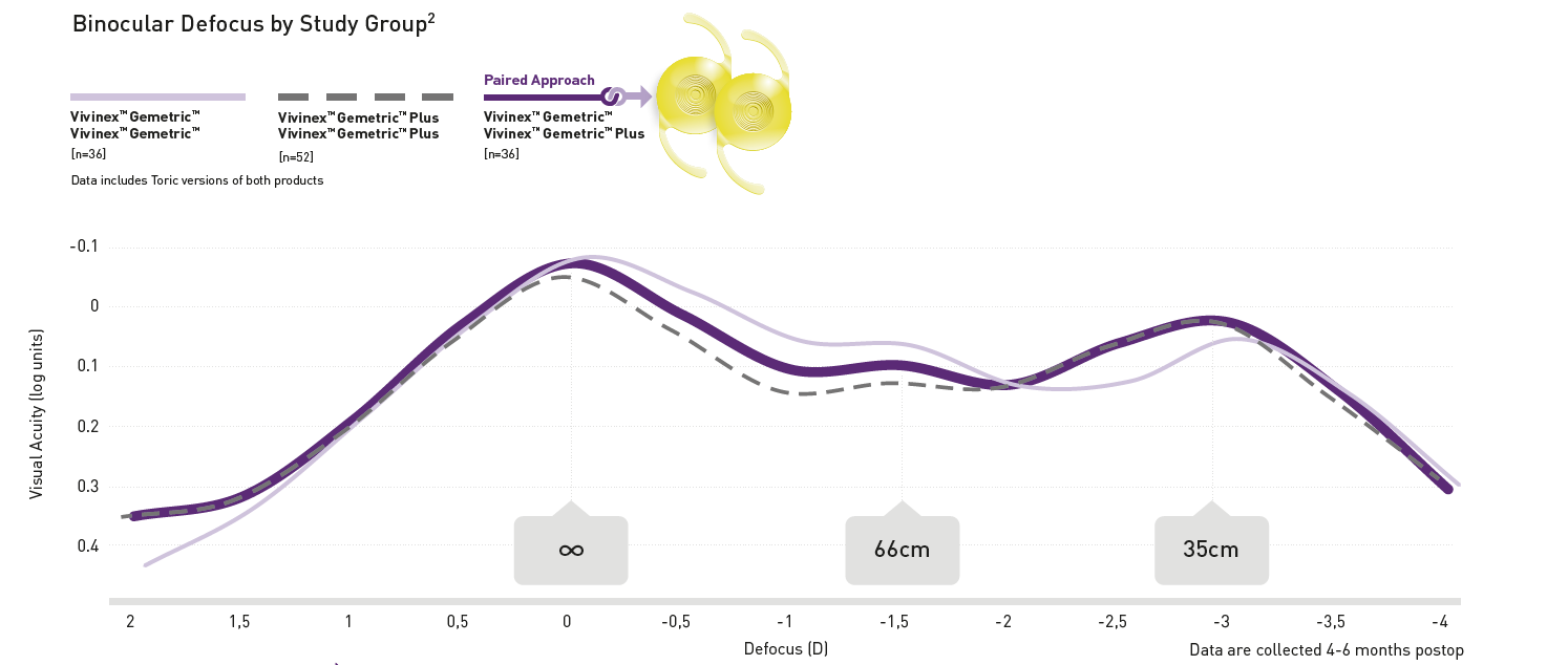
Die Vivinex Gemetric ermöglicht in der Ferndistanz ein exzellentes, im Intermediärbereich ein sehr gutes und in der Nähe ein gutes Sehen.
Die Vivinex Gemetric Plus ermöglicht in der Ferndistanz ein sehr gutes, im Intermediärbereich ein gutes und in der Nähe ein exzellentes Sehen.
Die zwei verschiedenen Gemetric-Designs können je nach Präferenz der Patientinnen und Patienten für die Ferne oder die Nähe aufeinander abgestimmt werden.
Die Vivinex Gemetric Plus ermöglicht in der Ferndistanz ein sehr gutes, im Intermediärbereich ein gutes und in der Nähe ein exzellentes Sehen.
Die zwei verschiedenen Gemetric-Designs können je nach Präferenz der Patientinnen und Patienten für die Ferne oder die Nähe aufeinander abgestimmt werden.

Ein breiter Sehbereich dank Pairing
Durch den Pairing-Ansatz können Patientinnen und Patienten einen noch grösseren Sehbereich angeboten werden. Beim Pairing wird eine Vivinex Gemetric mit einer Vivinex Gemetric Plus kombiniert.

Über 90 % Brillenunabhängigkeit
Eine postoperative Befragung hat ergeben, dass über 90 % der Patientinnen und Patienten 6 Monate nach der Katarakt-OP keine Brille mehr brauchen.

References:
1. HOYA data on file. CTM-23-029, HOYA Medical Singapore, Pte. Ltd, 2023 2. Miyata, A. et al. (2001): Clinical and experimental observation of glistening in acrylic intraocular lenses. In: Japanese journal of ophthalmology 45 (6), p. 564–569. 3. Auffarth et al. (2023) Randomized multicenter trial to assess posterior capsule opacification and glistenings in two hydrophobic acrylic intraocular lenses. Sci Rep 13, 2822. 4. Leydolt, C. et al. (2020): Posterior capsule opacification with two hydrophobic acrylic intraocular lenses: 3-year results of a randomized trial. In: American journal of ophthalmology 217 (9), p. 224-231. 5. Giacinto, C. et al. (2019): Surface properties of commercially available hydrophobic acrylic intraocular lenses: Comparative study. In: Journal of cataract and refractive surgery 45 (9), p. 1330–1334. 6. Werner, L. et al. (2019): Evaluation of clarity characteristics in a new hydrophobic acrylic IOL in comparison to commercially available IOLs. In: Journal of cataract and refractive surgery 45 (10), p. 1490–1497. 7. Matsushima, H. et al. (2006): Active oxygen processing for acrylic intraocular lenses to prevent posterior capsule opacification. In: Journal of cataract and refractive surgery 32 (6), p. 1035–1040. 8. Farukhi, A. et al. (2015): Evaluation of uveal and capsule biocompatibility of a single-piece hydrophobic acrylic intraocular lens with ultraviolet-ozone treatment on the posterior surface. In: Journal of cataract and refractive surgery 41 (5), p. 1081–1087. 9. Eldred, J. et al. (2019): An In Vitro Human Lens Capsular Bag Model Adopting a Graded Culture Regime to Assess Putative Impact of IOLs on PCO Formation. In: Investigative ophthalmology & visual science 60 (1), p. 113–122. 10. Nanavaty, M. et al. (2019): Edge profile of commercially available square-edged intraocular lenses: Part 2. In: Journal of cataract and refractive surgery 45 (6), p. 847–853. 11. Schartmueller, D. et al. (2019): True rotational stability of a single-piece hydrophobic intraocular lens. In: The British journal of ophthalmology 103 (2), p. 186–190. 12. Pérez-Merino, P.; Marcos, S. (2018): Effect of intraocular lens decentration on image quality tested in a custom model eye. In: Journal of cataract and refractive surgery 44(7), p. 889-896. 13. Miyata, A. et al. (2001): Clinical and experimental observation of glistening in acrylic intraocular lenses. In: Japanese journal of ophthalmology 45 (6), p. 564–569. 14. Pérez-Merino, P.; Marcos, S. (2018): Effect of intraocular lens decentration on image quality tested in a custom model eye. In: Journal of cataract and refractive surgery 44 (7), p. 889–896 15. Schartmuller, D. et al. (2019): True rotational stability of a single-piece hydrophobic intraocular lens. In: The British journal of ophthalmology 103 (2), p. 186–190.
